A/Prof George Kong, a glaucoma subspecialist and Dr Lukas Sahar have joined Dr Joseph San Laureano at Melbourne Eye Centre
A/Prof George kong
A/Prof. George Kong is a comprehensive ophthalmologist and Glaucoma subspecialist with a particular interest in cataract surgery, comprehensive glaucoma management, minimally invasive glaucoma surgery, and complex glaucoma caused by angle closure and inflammatory eye diseases. He also performs pterygium surgery, common eyelid surgeries and ICL refractive correction surgery.
A/Prof. Kong completed VCE in Melbourne finishing top of the state of Victoria in Mathematics. He then completed his medical degree (MBBS) from The University of Melbourne, Australia. He successfully completed his PhD in 2011 supported by National Health and Medical Research Council Postgraduate Training Scholarship. He was awarded the prestigious Eberhard Dodt Memorial Award from the International Society for Clinical Electrophysiology of Vision (ISCEV) for his work on retinal electrophysiology.
A/Prof. Kong completed RANZCO (Royal Australian and New Zealand College of Ophthalmologists) accredited training at the Royal Victorian Eye and Ear Hospital. Subsequently, he embarked on a subspecialty Glaucoma fellowship at The Royal Victorian Eye and Ear Hospital where he gained medical and surgical expertise in the treatment of glaucoma. He then travelled to Cambridge, United Kingdom, where he gained expertise in the advanced management of complex cataract and glaucoma conditions such as glaucoma caused by inflammatory eye diseases (uveitis) and children’s glaucoma. He has also trained in laser procedures for glaucoma and in new surgical techniques of Minimally Invasive Glaucoma Surgery.
A/Prof. Kong published over 50 international peer-reviewed journal articles, 5 book chapters and is a reviewer of several ophthalmology and optometry journals. He was instrumental in the development of the GONE (Glaucoma Optic Neuropathy Evaluation) website which has helped thousands of optometrists and junior ophthalmologists around the world improve their optic nerve examination techniques. He is a co-inventor of software for visual field testing software on iPad – Melbourne Rapid Fields. He is also the inventor of the Semi-open technique of XEN stent insertion. A/Prof. Kong was awarded the Gerard Crock Trophy in the RANZCO Annual Scientific Congress 2022 for his work on visual field home monitoring for glaucoma.
A/Prof. Kong speaks Mandarin and Cantonese fluently.
Dr Lukas Sahhar
Dr Lukas Sahhar is a comprehensive specialist ophthalmologist with additional training in complex cataract and refractive surgery.
He completed his medical degree at Monash University where he received several Dux awards as well as the Robert Power Prize in Surgery, the Neil Carson Prize in Medicine, the Monash Health Board of Management Prize, the Belinda Farrell Medicine Prize and the Hector Maclean Prize in Ophthalmology for Monash University. He received a scholarship to complete his Honours research degree with the Oxford Uehiro Centre for Practical Ethics at Oxford University and has also completed a Master of Public Health.
He undertook his specialist training in Melbourne at the Royal Victorian Eye and Ear Hospital, Alfred Hospital, Austin Hospital and Royal Children’s Hospital. As a registrar he was awarded the Australian and New Zealand Glaucoma Society Kath Holmes Prize. He undertook further subspecialist training as the Cataract Surgery Fellow at the Royal Victorian Eye and Ear Hospital in Melbourne where he gained expertise in managing complex cataract surgery cases and in using modern premium lens technologies.
Dr Sahhar is a fellow of the Royal Australian and New Zealand College of Ophthalmologists and a member of the Australasian Society of Cataract and Refractive Surgeons. He is a consultant ophthalmologist at the Royal Victorian Eye and Ear Hospital where he also teaches ophthalmologists in training.
Dr Sahhar manages conditions including:
Cataract with use of modern premium corrective lenses
Age related macular degeneration requiring injections
Diabetic eye disease
Glaucoma
Blepharoplasty
Pterygium
Eyelid entropion and ectropion
General ophthalmology
He customises his care and lens selection for the individual patient, offering monofocal, multifocal and extended depth of focus lenses tailored to each patient. He employs modern cataract surgery techniques including pain-free anaesthesia with drops (safe for patients on blood thinners), small incision surgery without sutures and eye patch-free recovery to promote early return of vision.
Dr Sahhar grew up in Warragul, Victoria and enjoys the outdoors and spending time with family and friends. He values connecting with his patients and providing friendly and personalised care.
Comparing Visual Field Tests: Humphrey Visual Field Analyzer vs. Virtual Reality Headsets
Our practice has recently moved to performing visual fields using the newly available VR headset.
Visual field tests are essential in eye care, helping ophthalmologists to detect and monitor conditions like glaucoma and neurological vision disorders. Traditionally, the Humphrey Visual Field Analyzer has been the go-to tool for these tests. However, newer technologies, such as virtual reality (VR) headsets offer an alternative that many patients find more comfortable and convenient. Let’s explore the key differences between these two methods and why VR headsets might be the future of visual field testing.
The Humphrey Visual Field Analyzer: The Traditional Approach
The Humphrey Visual Field Analyzer has been widelyused in eye clinics for years. This machine requires the patient to place their head inside a dome-like structure, resting their chin and forehead on supports to keep their head steady. Here’s what you can expect during a Humphrey visual field test:
– **Fixed Positioning:** Patients must hold their head still in a specific position, which can be uncomfortable, especially for those with neck or back problems. This can make the test challenging for some individuals, particularly those who have difficulty staying in one position for an extended period.
– **One Eye at a Time:** The Humphrey Analyzer tests each eye separately. To do this, a patch is placed over the eye not being tested. Patients often report discomfort from the patch, with some even saying it causes a distracting shadow, making the test less pleasant.
– **Technician Supervision:** During the test, a technician is required to monitor the patient closely. The technician ensures that the patient remains properly positioned and follows the test instructions. This human oversight is essential for accurate results but can make the experience feel somewhat clinical and rigid.
Virtual Reality Headsets: A Modern, Patient-Friendly Alternative
Virtual reality headsets offer a new way to perform visual field tests, with a focus on patient comfort and ease of use. Here’s how the experience with a VR headset differs:
– **Comfortable Positioning:** One of the most significant advantages of VR headsets is that they allow patients to take the test in a variety of positions. Whether sitting in a chair, reclining, or even lying down, patients can find a comfortable position that suits their needs. This flexibility is especially beneficial for those with neck or back issues.
– **Simultaneous Testing of Both Eyes:** Unlike the Humphrey Analyzer, the VR headset tests both eyes at the same time, eliminating the need for an eye patch. This not only makes the test more comfortable but also avoids the common complaint of a shadow caused by the patch, making the testing process more straightforward and less distracting.
– **Virtual Assistance and Eye Tracking:** The VR headset is equipped with a virtual assistant that guides patients through the entire process. The assistant explains how to do the test, monitors progress, and even offers encouragement along the way. This is a significant improvement over the traditional method, where a technician must supervise the test. Additionally, the VR headset includes advanced eye-tracking technology that helps minimize errors, leading to more accurate results.
– **Cloud-Based Data and Real-Time Monitoring:** Another innovative feature of VR headsets is their ability to store data in the cloud. This means that test results can be accessed from anywhere, and progress can be monitored in real-time through a web browser. This is particularly useful for both patients and doctors, as it allows for greater flexibility and convenience in managing eye health.
Patient Preference: VR Headsets Win
In trials conducted with patients, the response to VR headset testing has been overwhelmingly positive. Virtually all patients who have experienced both the Humphrey Analyzer and the VR headset have expressed a clear preference for the VR method. The comfort, convenience, and ease of use offered by VR technology make it a more patient-friendly option, and its innovative features provide a more modern approach to visual field testing.
Conclusion: The Future of Visual Field Testing
While the Humphrey Visual Field Analyzer remains a reliable and widely used tool, the introduction of VR headsets represents a significant advancement in eye care. By offering a more comfortable, convenient, and patient-centered approach, VR headsets are poised to become the preferred method for visual field testing in the future. Whether you’re concerned about comfort during the test, the need for more accurate results, or simply want a more streamlined experience, VR headsets offer an exciting new option that meets these needs and more.
This article follows on from my recent Zoom seminar which I recently posted, in which I presented current studies that have impact on the day to day management of glaucoma and cataract surgery.
In this seminar, which had over 200 registered attendees I looked at 4 studies, each of which will become the subject of an article
In this first instalment I discuss how I have changed my approach to cataract surgery following the recent FEMCAT study
Femtosecond, laser assisted cataract surgery (FLACS)
I first wrote about femtosecond Laser Assisted Cataract Surgery (FLACS) in May of 2013. In that article and many others that followed, I made the statement that I thought the FLACS was possibly the beginning of a new area in cataract surgery and would ultimately evolve to the point that it would be the new norm. The beginning of new era in cataract surgery with more and better to come.
I was so convinced that this was the way forward that I was even prepared to outrightly purchase a machine of my own. At over half a million dollars, that is no mean feat. In fact some did purchase their own, even sole practitioners. Thankfully I didn’t have to as a number of other ophthalmologists thought the same as me and so Victoria Parade Surgical Centre (VPSC) came to the party and bought one. They felt that they wanted to be an centre of excellence and therefore should have the latest technology. Since then, I have performed thousands of cases and in fact became the main surgeon, at VPSC performing FLACS.
In essence, these articles confirm what I was beginning to suspect and which many others were also beginning to suspect.
Findings of the Lancet article
The Lancet article is a Femtosecond laser-assisted versus phacoemulsification cataract surgery study (FEMCAT)
In this study there were total of 1476 patients, 704 had FLACS and 685 had conventional cataract surgery.
The study authors hypothesised that FLACS should provide superior outcomes over conventional cataract surgery because of its greater degree of precision and its superior ability to perform lens fragmentation, although at a greater cost.
The study looked at a number of endpoints:
1.Wether FLACS had more or less complications to conventional surgery
2.Wether visual outcomes were better or worse
3.Wether the ability to achieve the desired refraction was any different to conventional surgery
4. Wether astigmatism was correctable to a better degree
5.The incremental cost of the treatment (over a patient who did not have FLACS) and who has a good, non FLACS result
Their conclusion was that, to quote from the article, “Despite its advanced technology, femtosecond laser was not superior to phacoemulsification in cataract surgery and, with higher costs, did not provide and additional benefit over phacoemulsificaction for patients or healthcare systems.
So lets examine these points
Does FLACS have any serious complications
One of the main questions was wether FLACS resulted in a reduction in complications over standard cataract surgery. This question is probably the most frequently asked by patients. One of the findings about FLACS is that due to the lens fragmentation provided by the laser, less phaco energy Is required and so theoretically, this should lead to less endothelial cell loss and thus better outcomes. In practice, and from study findings, this has not really been borne out. My own impression is that softening a dense lens does not seem to make it easier to remove and I still find myself using phaco energy to break up the lens. Further, most lenses can be successfully pre-chopped, meaning that no sculpting is required. This is equivalent to the laser breaking up the lens, also requiring no sculpting
The study concluded that FLACS was as safe as conventional surgery but no safer. Further, there did seem to be an increased tendency for anterior capsular tears, which has also been my experience.
Are Visual outcomes better with FLACS
By this, the question is wether the best corrected vision (with or without glasses) was any different to standard surgery
The findings were that FLACS was not inferior but also no superior. This means that patients having FLACS were just as likely to achieve their best corrected vision as those patients having conventional surgery.
Does FLACS achieve desired refraction any better
One of the problems with standard cataract surgery is achieving the desired refractive outcome unaided. That is to say, that if the goal is emmetropia then post operatively, patients ideally should be within 0.5 Diopters (and ideally within 0.25) of Plano to enable good distance vision without glasses.
In practice, most surgeons can only achieve this 85% of the time. This leaves a 15% group that will still require distance correction or in need of a further procedure to achieve that goal.
With FLACS, the hope was that with greater precision, would come greater refractive predictability. In reality, this has not been the case.
It would seem that although a capsulorhexis performed with a laser is perfectly round and perfectly sized compared to a manual rhexis, this has not resulted in any great benefit. Theoretically a rhexis that is too large will allow forward vaulting of the intraocular lens and therefore a myopic outcome and conversely, a rhexis that is too small will result in a more posterior position of the lens and therefore, potentially a hyperopic outcome.
Refractive outcomes with FLACS have not been found to be any better than with standard surgery.
Similarly, patients with astigmatism are no better of with FLACS
Cost of laser assisted cataract surgery
Laser cataract surgery is unquestionably expensive and the conclusion of the study is that it is not cost effective
So who should have FLACS?
The one area where laser assisted surgery is potentially better is in patients who have an advanced, intumescent cataract where the risk of an anterior scapular tear is high (Argentinian flag sign)
As the laser performs the capsulorhexis in under one second, the risk of a tear is greatly reduced although not entirely eliminated.
Patients with loose zonules such as those with PXF may also benefit as the rhexis is created without further zonular stress.
A manual capsulorhexis with result in further dehiscence of the lens due to zonular tearing increasing the risk of a dropped lens or lens dislocation
Patients with endothelial dystrophy such as as Fuch’s may also be better of as there may be less endothelial cell loss but even here, with pre-chopping there may be little benefit.
How I have changed my approach to cataract surgery
I no longer routinely offer FLACS as it is not cost effective.
I now offer it to patients who have significant PXF and who appear to have loose zonules
If a patient has an advanced, intumescent cataract I recommend it as a means of minimising the risk of a capsular tear.
Lastly, if a patient has obvious, significant zonular laxity for example after trauma or in patients with Marfan’s, I also offer it.
If the cost of FLACS comes down with time, which I do not believe will happen then I will offer more widely but at present, the cost is prohibitive and not justified
I recently gave a presentation over Zoom discussing a number of recently published journal articles that have altered how we manage patients with glaucoma and how I have altered my approach to laser assisted cataract surgery.
Two hundred optometrists registered for the meeting
In a future post, I will discuss these findings in greater detail
Below is this presentation which is in three parts with questions after each part.
The first part discusses the role of SLT in managing open angle glaucoma and is based on the recently published LiGHT study, the second part discusses the ZAP and Eagle studies looking at the role of iridotomies and lens surgery in managing narrow angles and the last part discusses the Femcat and related studies and I talk about how this has influenced my approach to cataract surgery
Melbourne eye centre has a new website which went live on Wednesday the 8th of January 2020
The old site served us well for over 17 years but was in need of modernising.
This new website makes it easier to find information about our laser eye surgery services.
We will be updating the site regularly with interesting topics on in the areas in which we specialise which is cataract surgery, refractive surgery and glaucoma surgery
As you can see, we use the term progressive eye care as our focus is on being the first and the best with the most current techniques in our areas of expertise


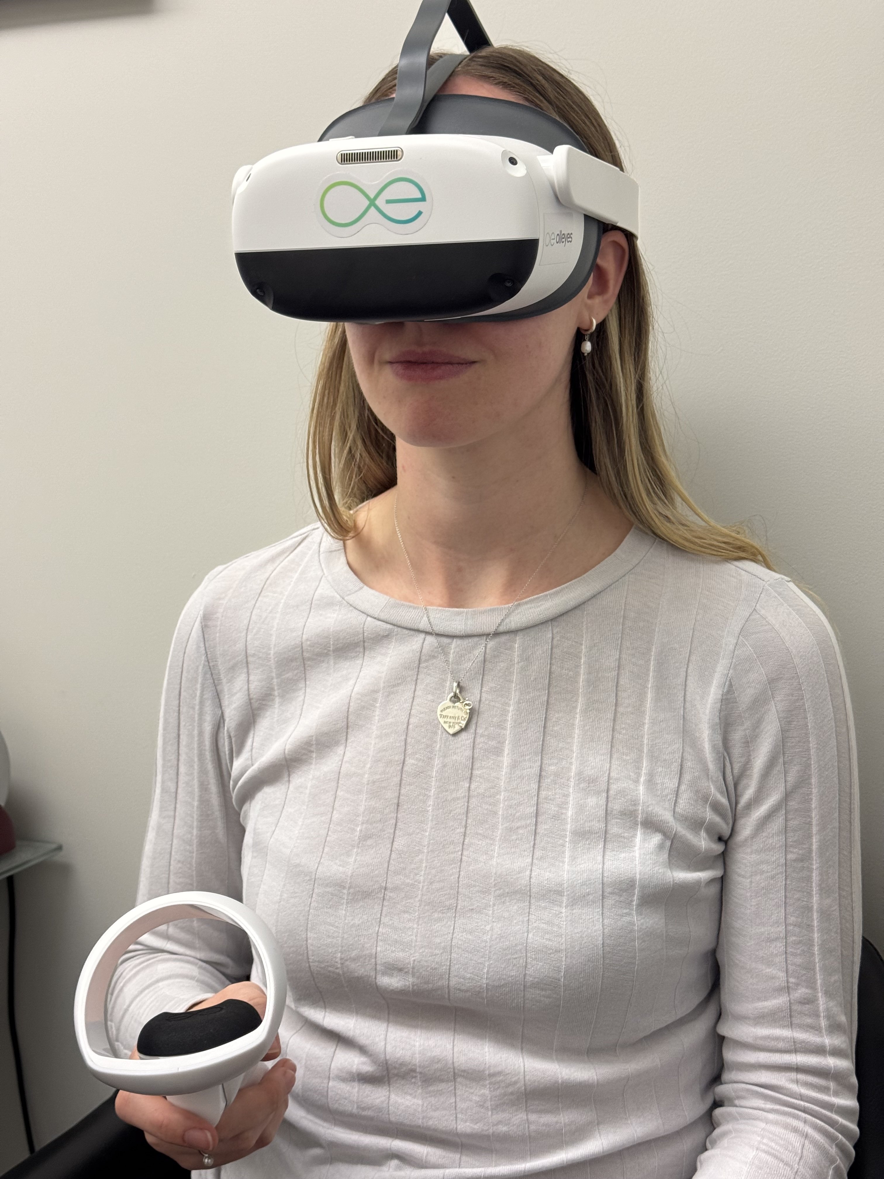 Virtual reality headsets offer a new way to perform visual field tests, with a focus on patient comfort and ease of use. Here’s how the experience with a VR headset differs:


